
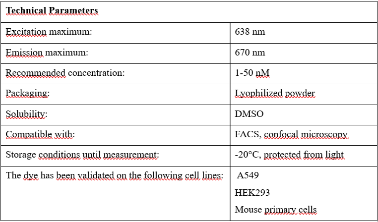
BioxML Red
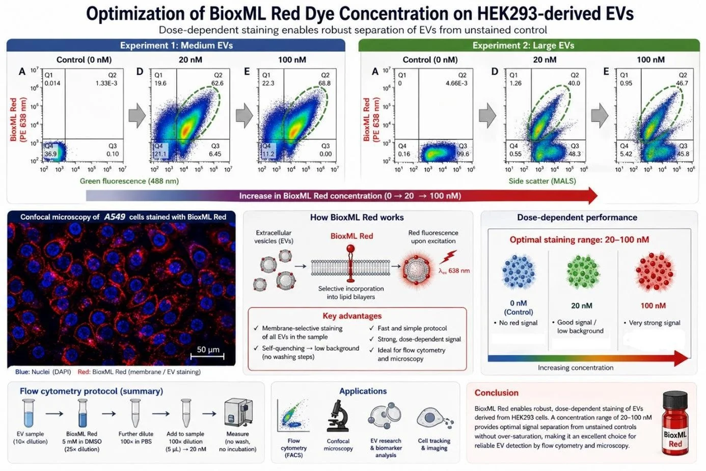
BioxML Red is a red fluorescent dye specifically developed by Bioxol Ltd. for the reliable and specific labeling of extracellular vesicles as well as cell membranes. The dye selectively incorporates into lipid bilayers, where, upon excitation with light of an appropriate wavelength, it emits a red fluorescent signal. It is suitable for both flow cytometry and confocal microscopy applications. The Institute of Genetics, Cell and Immunobiology at Semmelweis University is already actively using it in various experimental studies.
BioxML Red addresses common challenges encountered in the study of extracellular vesicles that hinder their detectability and the reproducibility of their measurement:
• Non-specific staining
• Background signal caused by aggregation
• Lengthy or sensitive antibody staining protocols
• Difficulties arising from the lack of knowledge of EV markers
• Weak fluorescence signal due to the micrometer-scale size of EVs
What BioxML Red provides:
• Ensures membrane-selective localization, staining all EVs present in the sample, thereby enabling comprehensive quantitative analysis.
• Self-quenching capability, allowing low background without the need for washing steps.
• A fast and simple protocol that shortens the process of EV detection in flow cytometry.
Protocol for EV detection by flow cytometry:
• The sample containing EVs isolated and purified from the conditioned medium of HEK293 cells was first diluted 10×, and a final 50× dilution was used for the experiment.
• A stock solution of BioxML Red (5 mM) was diluted 25× in DMSO under sterile conditions (biosafety cabinet).
• In the next step, it was further diluted 100× in PBS.
• For the experiment, the dye was added to the samples at a final 100× dilution, corresponding to 5 µL, resulting in a final dye concentration of 20 nM.
• Based on experience gained during protocol development, no additional washing steps or incubation time are required.
Notes:
+ The PBS solution used in the experiments was prepared by diluting 5 ml of 10× PBS (SE, Egyetemi Gyógyszertár ) with 45 ml of Milli-Q water (Merck) in a Falcon tube, followed by filtration through a 0.2 µm filter ( Millipore) to avoid physical contamination.
+ Successful EV staining was confirmed using control samples treated with detergent lysis at appropriate concentrations.
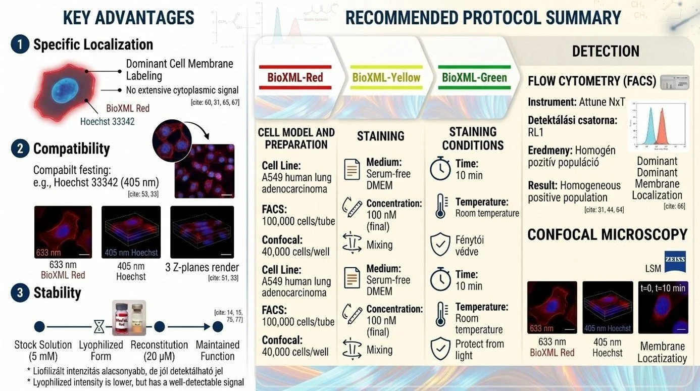
The product is an ideal choice for:
• Flow cytometry (FACS) applications
• Confocal microscopy
• EV research and biomarker analysis
• Studying cell membrane dynamics
• Cell tracking and imaging
Related articles:
The BioxML Red is part of the Bioxol dye family, and several scientific publications on its application are expected to appear in the near future:
Koncz, A., Turiák, L., Németh, K., Lenzinger, D., Bárkai, T., Lőrincz, P., Zelenyánszki, H., Vukman,
K. V., Buzás, E. I., & Visnovitz, T. (2023). Endoplasmin Is a Hypoxia-Inducible Endoplasmic
Reticulum-Derived Cargo of Extracellular Vesicles Released by Cardiac Cell
Lines. Membranes, 13(4), 431. https://doi.org/10.3390/membranes13040431
Several scientific publications are already available on another member of the dye family, BioxML Yellow:
1. Benke, M., Zeöld, A., Kittel, Á., Khamari, D., Hritz, I., Horváth, M., Keczer, B., Borka, K., Szücs, Á., & Wiener, Z. (2023). MiR-200b categorizes patients into pancreas cystic lesion subgroups with different malignant potential. Scientific reports, 13(1), 19820. https://doi.org/10.1038/s41598-02347129-1
2. Lenzinger, D., Lankovics, L., Dudás, I., Bárkai, T., Szász, Z., Vukman, K. V., Fletcher, K., Csomos, A., Mucsi, Z., Bugyik, E., Cserép, C., Dénes, Á., Bősze, Sz., Buzás, E. I., & Visnovitz, T. (2025). Stress-induced switch in small extracellular vesicle secretion: From constitutive “torn bag mechanism” to exocytosis. bioRxiv. https://doi.org/10.1101/2025.10.29.685290